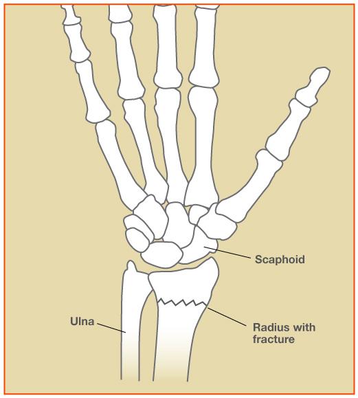
The wrist is composed of eight small bones and the two forearm bones, the radius and ulna. The complex anatomy allows the wrist to move in so many directions and is why our wrist can bend and straighten, move side-to-side, and rotate. Restoration of normal anatomy is the best way to regain function after a fracture or break of the wrist.
While any combination or single bone can be fractured, there are a couple fractures that are most common. A couple of these common fractures are distal radius fractures, and scaphoid fractures.
Click here to learn more about distal radius fractures.
The first step in evaluating a fracture is to evaluate the bones using an x-ray. X-rays allow me to see the alignment of your wrist bones. If the bones are still well aligned, treatment may be possible with a period of rest in a splint or cast. On occasion, an MRI or CT scan is needed to obtain a closer look at the bones.
Treatment for Wrist Fractures
If alignment of the bones is not an issue, a splint or cast can be worn to keep the bones from displacing. Routine x-rays will be taken to ensure the bone is healing well and hasn’t moved out of place.
Wrist Fracture Surgery
If the bones need to be re-aligned, surgery is recommended to restore the complex anatomy of the wrist. There are a variety of ways to hold the fractured bone in place, depending on the fracture. Some of these include plates, screws, wires, or rods. The goal of surgery is to restore the normal anatomy of the wrist. This also allows early motion to prevent stiffness.
Diagrams modified from ASSH (www.assh.org)